TECHNOLOGY
Reading the brain with light.
What the signal is
When neurons become active, they briefly change shape. Ion and water movement across their membranes makes the cells swell by a small amount, and the surrounding tissue changes with it. Swelling changes how the tissue scatters and absorbs near-infrared light.
We shine light in, measure what comes back, and the scattering change mirrors the electrical event that caused it, leading to a signal 1000x faster than ones that measure blood flow, like fNIRS and fMRI. The signal is called the Fast Optical Signal or Event-Related Optical Signal.
How it differs from fNIRS and EEG
The useful distinction is not how fast each one is — it is what physical quantity each one is actually sensitive to.
| Method | What it measures | Timing | Main limitation |
|---|---|---|---|
| EEG | Summed electrical potentials at the scalp | Milliseconds | Low spatial accuracy |
| fNIRS | Hemoglobin concentration, via absorption | Seconds | Measures blood, not neuronal activity |
| FOS | Changes in light scattering from neuronal activity | Milliseconds | Low signal-to-noise — the part we're changing |
fNIRS and FOS are both optical, but they measure very different things. FOS is an optical measurement of neuronal events.
How the measurement works
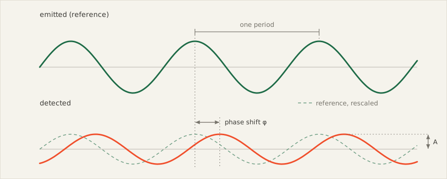
A frequency-domain instrument modulates the light source at radio frequency rather than shining a constant beam. When neurons are active, the tissue becomes more transparent and the average path of the photons becomes deeper. This means that on average, photons take longer to reach the detector. With a frequency-domain instrument, we measure this change in photon time of flight as a shift in phase.
What is hard about it
A note on what we do not publish
We have patent work in flight, so this page describes the public science and the shape of the problem rather than our approach to it. What we can say publicly is that on some of these problems, we have moved further than the published state of the art.
WHERE THIS GOES
Next up
Prove the hardware on optical phantoms before trusting it on a person. Done means a scorecard we would show a skeptical reviewer, with a noise floor we can defend and results that repeat across builds.
Record a known, well-replicated response in real subjects — the kind of result the FOS literature already contains, so that agreement means something. Done means matching published effects on our own hardware.
Move from detecting that something happened to identifying what. Done means single-trial performance good enough that the measurement is useful in minutes.
Safety
The instrument emits near-infrared light at levels well below established exposure limits for skin and tissue. A useful comparison: more photons enter your skull on a sunny afternoon than our instrument delivers. Nothing is implanted, nothing except light penetrates the skin, and a session can be stopped at any moment by taking the headgear off.
Selected references
The FOS literature is public, and reading it is the fastest way to decide whether you believe any of this. Three to start with; the full list, with the sources behind every figure on this site, is on the research page.
- Shades of gray matter: noninvasive optical images of human brain responses during visual stimulation.
Gratton, G., Corballis, P. M., Cho, E., Fabiani, M., & Hood, D. C. (1995).
Psychophysiology, 32(5), 505–509. - Fast optical imaging of human brain function.
Gratton, G., & Fabiani, M. (2010).
Frontiers in Human Neuroscience, 4, 52. - The fast optical signal — robust or elusive when non-invasively measured in the human adult?
Steinbrink, J., Kempf, F. C. D., Villringer, A., & Obrig, H. (2005).
NeuroImage, 26(4), 996–1008.