Putting FOS on the Map
Fast Optical Signal (FOS) is now on the BrainMind BCI Universe Map.

There's a new entry in the BCI Universe. BrainMind's 2026 map of brain-computer interface technologies now includes Fast Optical Signal (FOS). It sits on the noninvasive side of the map, on the (new!) Optical branch, right next to fNIRS.
That might sound like a small thing, but FOS has spent three decades mostly inside a handful of academic labs, and maps like this are where students, founders, clinicians, and funders go to get their bearings in a crowded field.
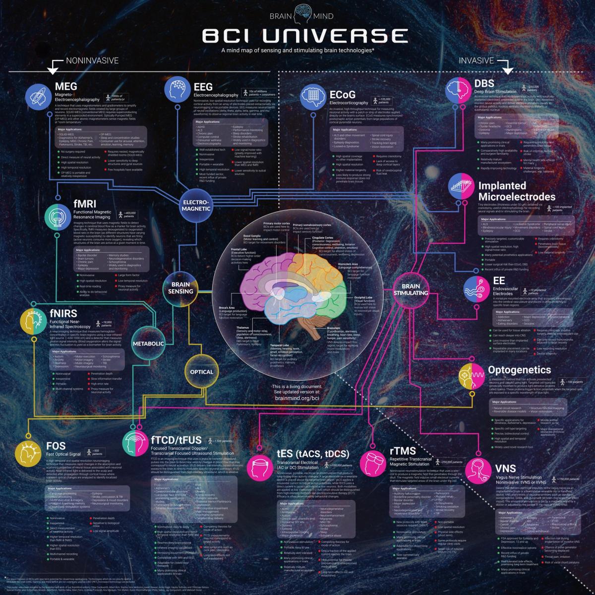
BrainMind's BCI Universe 2026. FOS appears at lower left, on the Optical branch. See the living version at brainmind.org/bci.
Who BrainMind Is
BrainMind is a community of scientists, engineers, clinicians, entrepreneurs, investors, and philanthropists. Its mission is to "support and cultivate the most important and impactful ideas in brain science," with special attention to areas that are under-supported relative to their importance. Founder Michael McCullough describes the gap it fills: grants fund basic lab research, venture capital funds profitable companies, and many high-impact ideas fall in between.
The BCI Universe is one of the ways BrainMind maps the field. It's a single poster – "a mind map of sensing and stimulating brain technologies" – covering 14 of them, from EEG and fMRI to deep brain stimulation and optogenetics. Each gets a plain-language description, its major applications, its strengths and weaknesses, and a rough count of patients. The focus is on BCIs with near-term potential for closed-loop applications, and it's a living document, kept current at brainmind.org/bci.
What BrainMind Says About FOS
Here is BrainMind's entry for FOS, in full:
FOS – Fast Optical Signal (~500 patients)
A high temporal and spatial resolution neuroimaging technique that measures rapid changes in the absorption and scattering properties of neural tissue associated with neuronal activity. Near-infrared light is delivered to the scalp and detected after propagation through cortical tissue where transient optical changes are analyzed to identify localized brain activity.
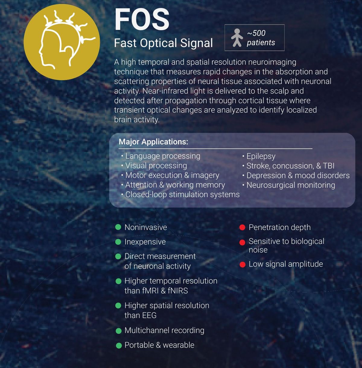
The FOS entry on the BCI Universe 2026. Courtesy of BrainMind.
Major applications:
- Language processing
- Visual processing
- Motor execution & imagery
- Attention & working memory
- Closed-loop stimulation systems
- Epilepsy
- Stroke, concussion, & TBI
- Depression & mood disorders
- Neurosurgical monitoring
Strengths:
- Noninvasive
- Inexpensive
- Direct measurement of neuronal activity
- Higher temporal resolution than fMRI & fNIRS
- Higher spatial resolution than EEG
- Multichannel recording
- Portable & wearable
Challenges:
- Penetration depth
- Sensitivity to biological noise
- Low signal amplitude
That's a fair scorecard; the strengths are why we started Brill, and the challenges are what we’re working to improve every day.
FOS, In Plain English
When neurons fire, they physically change. It’s an impossibly tiny change, but as those neurons swell and shift ever so slightly, the way light scatters through that tissue changes as well. Essentially in areas where populations of your neurons are active, the light goes deeper and is delayed by picoseconds. FOS measures that change.
The way it works in practice is by taking harmless near-infrared light, similar to your Apple Watch, but turned bright and dim very fast so it’s modulated like a wave, and when it’s pressed to a person’s head, a small fraction of it passes through the skull, reaches the brain's outer layer (cortex), and scatters back out to a detector a few centimeters away. When the neurons along that path become active, we see a slight delay in that waveform and measure that shift. Each measurement occurs within milliseconds, but the delay or “shift” in the light is measured in picoseconds – a trillionth of a second!
Most noninvasive brain imaging doesn't watch neurons at all. fMRI and fNIRS measure changes in blood flow in the brain, looking for the rush of oxygenated blood that arrives a few seconds after neurons fire. fMRI and other “hemodynamic” methods looking at blood flow can be precise, but they’re slow – on the order of 3-5 seconds, which is 1000x slower than FOS, which measures in windows of 3-5 milliseconds. Electrical methods like EEG picks up fast electrical activity, but it blurs as it passes through the skull, and its much less spatially precise than FOS. With EEG you get the when without much of the where. FOS aims for both: millimeters and milliseconds.
Where FOS Came From
The idea that active nerves change how they handle light is older than the personal computer. In 1968, Larry Cohen, Richard Keynes, and Bertil Hille reported in Nature that nerve fibers change how they scatter light while firing. But that was in isolated nerves on a lab bench, not through a living human skull.
That next leap came in 1995: Gabriele Gratton and Monica Fabiani, with their colleagues, published "Shades of Gray Matter", showing noninvasive optical images of the human visual cortex responding to stimulation. A demonstration in motor cortex followed that same year. They named their method EROS, the event-related optical signal, and you'll see FOS and EROS used almost interchangeably in the literature.
Gratton and Fabiani have spent the three decades since refining it, today from the Cognitive Neuroimaging Laboratory they direct at the University of Illinois Urbana-Champaign's Beckman Institute. Their work built the evidence that FOS is real, replicable, and useful for studying attention, memory, aging, and more. What FOS never had was a dedicated device built for it, or a company to take it out of the lab.
Where Brill Comes In
Built on the heritage of the UIUC’s CNL, and ISS, Inc. who made the first commercial device capable of detecting FOS, Brill Neurotech is building the first dedicated device for FOS. Rather than adapting general-purpose optical equipment, we're designing ours from the ground up around one job: capturing a very small, very fast signal, reliably, portably and outside of a specialist lab.
That means taking BrainMind's list of challenges head-on. Low signal amplitude and biological noise are big engineering problems. They come down to light sources, detectors, electronics, and signal processing, and that's where most of our effort goes. Depth is physics: FOS reads the brain's outer layer, no more than a few centimeters beneath the scalp. But that outer layer, the cortex, is where much of language, vision, movement, and attention takes place. The cortex is where humans interact with the world, which is perfect for Brain-Computer Interfaces.
Gabriele Gratton serves on our advisory board; our bench work happens in Champaign, just down the road from the Beckman Institute. And our CEO, Mehmet Günal, has worked at both UIUC’s CNL as well as ISS, and is among those BrainMind thanked on the poster.
What's Next
This is the first post on our new site, so we kept it short. Next, we'll publish a deeper look at FOS: how it works, what the research shows, and why we think it's one of the most under-explored signals in neuroscience. By our count of the published literature, there are about 136 FOS studies, compared with nearly 20,000 for fNIRS and 542k+ for EEG. There's a lot of room to grow.
Thank you to BrainMind and Diana Seville, plus its editors – Chloe Duckworth, Albert Kim, Sophia Vera Verduzco, Isaiah Nawaz, Anna Riggs, Hayley Sullivan, and Chinmayi Balusu – for putting FOS on the map. You can explore the full BCI Universe at brainmind.org/bci.